Atlas: Organ and Tissue Anatomy

This webpage introduces atlases of Xenopus, including books, papers, and external web content that have been released independently of our project. On clicking the icons below, you will be redirected to external websites where more details can be found.
An atlas originally means refers to "a book of maps." In biology, an atlas is similar to a book of maps that contain basic biological information of an organism, such as the positions and names of the cells, tissues, and organs. We are considering creation of an atlas of Xenopus tropicalis and publication of it on this website.
G. Bernardini, M. Prati, E. Bonetti, G. Scari (1999)
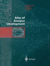
Images of tissue sections etc. made from early Xenopus laevis embryos. You need to buy this book if you want to read the book.
(9.9.2019 update)

Color Atlas of Xenopus laevis Histology
Allan F. Wiechmann, Celeste E. Wirsig-Wiechmann (2003)
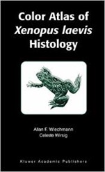
Images of tissue sections etc. made from matured Xenopus laevis frog. You need to buy this book if you want to read the book.
(9.9.2019 update)

Digital Xenopus Atlases in Xenbase webpage. Many line-drawings and images excerpted from papers and books are put on Xenbase webpage. Some contents require Java.
(9.9.2019 update)
Tissue section-Images of thyroid and gonad during embryonic developmental stage (22) to froglet stage (66).
(1.15.2018 update)
Three-dimensional morphology of inner ear development in Xenopus laevis.(論文・英語 / PubMed)
Bever MM, Jean YY, Fekete DM. (2003)
The three-dimensional morphology of the membranous labyrinth of Xenopus laevis is presented from embryonic through late tadpole development (stages 28 to 52, inclusive).
(1.15.2018 update)


Books
It is possible to browse sample pages on Google Books, before buying the following books.
Web Contents
-
Japanese / English
-
Atlas
Copyright Ⓒ NBRP X. tropicalis. All rights reserved.